
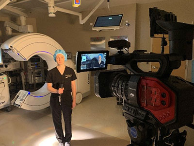
Southern Hills Hospital and Medical Center for the first time used a new imaging system that will assist surgeons in navigation techniques and help the hospital expand and enhance surgical procedures.
The technology is called the O-arm Imaging System, the equipment provides complete, multi-dimensional surgical imaging. It provides surgeons with real-time, 3-D images, as well as multiplane, 2-D and fluoroscopic imaging.
Coupling this system with other computer-assisted navigation equipment, Southern Hills Hospital surgeons can perform procedures with a higher rate of accuracy, potentially allowing them to successfully perform even more difficult and complex surgeries.
"Adding this surgical imaging and navigation system allows us to further streamline operating room efficiency and improve patient outcomes," says Alexis Mussi, CEO of Southern Hills Hospital and Medical Center.
According to Dr. Angela Palmer, a neurosurgeon with the hospital, complex spinal deformities make it very difficult to work on a patient. Using the more traditional X-ray equipment, surgeons had to take several, lesser-quality images of the patient, often having to move the patient and equipment into different positions to get the best view of the area of the spine they were working on.
"Traditionally, we use anatomic landmarks to place screws in the spine, but each person's anatomy is different. There are differences in bone diameter, density, and shape," explains Palmer.
“Because we are working within a very narrow channel of bone, there can be no margin of error. Computer-assisted surgical navigation will help us become even more accurate when implanting devices in the spine.”
In addition to its use on patients with complex spinal anatomy, the imaging system will be used in more routine, minimally invasive surgical procedures, providing for smaller incisions and a reduction in surgery time.
Potential benefits
Among the potential benefits of this technology are:
- Smaller incisions, resulting in less pain, quicker recovery times, and a decreased chance of infection or other complications.
- Fast access to multidimensional images. Images are produced in approximately 30 seconds, reducing overall time in surgery and reducing the number of times patients are under anesthesia.
- Verification of implant hardware placement. Surgeons can view a patient's anatomy, verify placement and make any adjustments before the patient leaves the operating room.
- Visualization of implant hardware placement can potentially reduce the need for follow-up surgery.